

Key Takeaways
- Fascia, tendons, and ligaments have far fewer blood vessels than muscle, slowing their repair significantly.
- Connective tissue remodeling can take months to years, even when pain has subsided.
- Most generic recovery advice targets muscle, leaving connective tissue needs underserved.
- Adequate protein intake, sleep, and progressive loading all support connective tissue health.
- Returning to full training intensity before connective tissue has adapted is a leading cause of overuse injury.
- Consulting a healthcare professional is essential for any suspected ligament or tendon injury.
Connective Tissue (Fascia, Tendons & Ligaments)
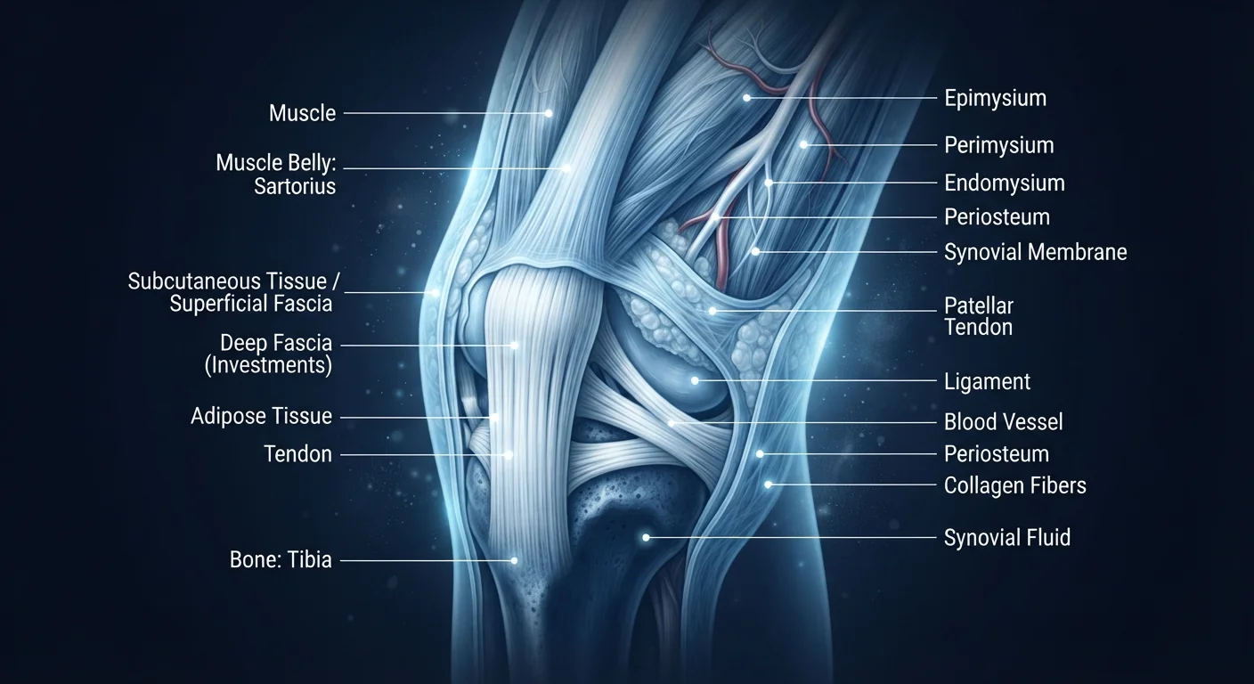
Fascia, tendons, and ligaments are dense connective tissues that hold the body together, transmit force, and stabilize joints. Unlike muscle, they contain relatively few blood vessels and cells, which means they receive less oxygen and nutrients - and repair themselves considerably more slowly after stress or injury.
These tissues are primarily composed of type I collagen fibers and have a low cellularity (fibroblast density), resulting in a limited regenerative capacity compared to skeletal muscle tissue.
The Tissues Most Recovery Plans Ignore
When you feel sore after a hard training session, the discomfort you're sensing is mostly muscular. Delayed-onset muscle soreness (DOMS) is familiar, measurable, and - crucially - temporary. Within 48 to 72 hours, muscle tissue has typically begun meaningful repair. But beneath that surface-level soreness, another set of structures is quietly absorbing stress and adapting on a much longer timeline: your connective tissues.
Fascia, tendons, and ligaments don't announce themselves with the same sharp post-workout signals. They adapt slowly, fail silently, and - when overloaded without adequate recovery - become the source of the nagging, persistent injuries that derail long-term training. Understanding how these tissues work is foundational to building physical resilience that lasts. See what actually happens to your muscles after a hard workout for a contrast with how muscle tissue responds to the same stress.
What Each Tissue Does - and Why It's Slow to Heal
Fascia is a continuous, three-dimensional web of fibrous tissue that encases and connects muscles, bones, nerves, and organs. Think of it as the body's internal scaffolding - it transmits mechanical force and helps coordinate movement across regions. When fascia becomes restricted through injury, chronic tension, or inactivity, movement quality suffers throughout the kinetic chain.
Tendons connect muscle to bone and transfer the force a contracting muscle generates into skeletal movement. They are built for tensile strength, not flexibility, and under repeated loading they accumulate micro-damage that must be cleared and replaced by new collagen. This process - tendon remodeling - can take months, not days.
Ligaments connect bone to bone, providing passive joint stability. They are especially vulnerable during sudden changes of direction or joint forces that exceed their structural limits. Because ligaments have limited vascularity (blood supply), the healing process is slow and the risk of incomplete recovery is real.
~10×
Difference in blood vessel density: muscle vs. tendon
Research in tendon biology consistently identifies low vascularity as the primary limiting factor in tendon healing speed compared to skeletal muscle.
6-12 months
Typical ligament remodeling timeline
Orthopedic research indicates that even after pain resolves, ligament collagen remodeling continues for six months to over a year post-injury.
30%
Decline in collagen synthesis rate with aging
Studies estimate collagen production declines meaningfully from early adulthood, contributing to increased connective tissue injury risk in older adults.
The common thread across all three tissues is low blood vessel density. Where muscle receives a rich, responsive blood supply that accelerates nutrient delivery and waste removal, connective tissue operates in a comparatively sparse vascular environment. Fewer vessels mean slower healing - even when everything else is optimized.
The Mismatch Between Muscle Recovery and Connective Tissue Recovery
Here's where many exercisers run into trouble: their muscles recover faster than their connective tissues, creating a false sense of readiness. By the time DOMS has resolved and an athlete feels capable of training hard again, the tendons and fascial structures supporting those muscles may still be mid-repair.
This mismatch is especially pronounced during periods of rapid training progression - when volume or intensity jumps faster than connective tissue can adapt. The result is often a classic overuse injury: Achilles tendinopathy, patellar tendon pain, plantar fasciitis, or iliotibial band syndrome. Each of these is a connective tissue failure driven not by a single traumatic event, but by accumulated load that outpaced recovery capacity.
Understanding the phases of physical recovery - including the slower remodeling phase - helps contextualize why connective tissue demands a longer timeline and more deliberate programming.
What Supports Connective Tissue Recovery
While connective tissue heals more slowly than muscle, it does respond to the right inputs. Several evidence-informed factors support the process:
- Adequate protein intake: Collagen is a protein, and its synthesis depends on sufficient dietary amino acid availability. General guidance for active adults emphasizes meeting overall protein needs consistently - not just around workouts.
- Quality sleep: Growth hormone - released primarily during deep sleep - plays a role in tissue repair across the body, including connective structures. Chronic sleep restriction impairs this process. Learn more about why recovery slows with age and the biological factors involved.
- Progressive loading: Tendons and ligaments strengthen in response to mechanical stress - but only when that stress is applied gradually and with adequate rest between sessions. Sudden spikes in training load are a primary injury trigger.
- Circulation-supporting movement: While connective tissue is less vascular than muscle, gentle movement promotes synovial fluid distribution and nutrient diffusion into dense tissues. Complete inactivity is rarely optimal. The choice between active recovery and complete rest matters here.
This article is for general informational purposes only and does not constitute medical advice. Always consult a qualified healthcare professional for evaluation of any injury, pain, or before making significant changes to your training or recovery approach.