
Skin Cancer Screening: What Dermatologists Look for During a Full-Body Check

Key Takeaways
- A full-body skin exam covers every surface, including scalp, nail beds, and areas not exposed to the sun.
- Dermatologists use the ABCDE criteria to evaluate moles and lesions for potential malignancy.
- People with fair skin, a personal or family history of skin cancer, or significant sun exposure benefit most from regular exams.
- Early detection dramatically improves outcomes for all three major types of skin cancer.
- Preparing properly — removing nail polish, wearing hair loose — helps ensure nothing is missed.
What you will need
Why Skin Cancer Screening Matters
Skin cancer is the most commonly diagnosed cancer in the United States, yet it is also among the most treatable when caught early. According to the American Academy of Dermatology, one in five Americans will develop skin cancer during their lifetime. Despite those numbers, many people have never had a professional full-body skin exam.
Unlike many internal cancers, skin cancer is visible — which is both an advantage and the reason a trained clinician's eye is so valuable. Dermatologists are equipped to distinguish benign spots from early warning signs that might look innocuous to an untrained observer. Early detection genuinely changes outcomes: melanoma detected at stage I has a five-year survival rate above 98%, compared with roughly 30% when found at stage IV.
If you want context on where skin checks fit alongside other preventive visits, see our overview of preventive screenings by life stage.
Practice Monthly Self-Exams Between Visits
Professional exams are valuable, but your own eyes are your first line of defense between appointments. Using good lighting and a full-length mirror — plus a hand mirror for hard-to-see areas — do a monthly review of your skin from head to toe. Note any new spots or changes to existing ones, and report anything unusual to your dermatologist promptly rather than waiting for your next scheduled visit.
Who Should Prioritize Regular Skin Exams
While anyone can develop skin cancer, certain factors raise individual risk and make regular professional exams especially important:
- Fair or light skin, light eyes, or red/blonde hair — less melanin means reduced natural UV protection.
- History of sunburns, especially blistering burns in childhood
- Personal or family history of skin cancer or atypical moles
- Significant cumulative sun exposure, including outdoor occupations or living at high altitude
- History of tanning bed use
- Immunosuppression from medications or medical conditions
That said, skin cancer occurs across all skin tones. Darker complexions tend to develop melanoma in less sun-exposed areas — palms, soles, under the nails — where lesions are easier to overlook. Annual or more frequent exams are worth discussing with a dermatologist regardless of skin tone if any risk factors apply.
Skin exams are one of several screenings that patients may not always think to request proactively. Our article on screenings your doctor may not always mention first explores this gap further.
How to Prepare for Your Appointment
A little preparation ensures the dermatologist can examine every surface of skin without obstruction.
What you will need
What the Dermatologist Evaluates: Step by Step
Understanding what happens during the exam can ease any anxiety about the process.
Check-In and Medical History Review
The appointment begins with a brief intake review. The dermatologist or a clinical assistant will ask about your personal and family history of skin cancer, any new or changing spots you've noticed, sun exposure habits, and medications or conditions that may affect skin health. Be thorough — even details that seem minor may be clinically relevant.
Full-Body Disrobing and Gowning
You will be asked to undress and put on a medical gown. A complete skin exam covers the entire body surface — front, back, and sides. Remaining areas covered mean areas unexamined, which is why full disrobing (to undergarments, at minimum) is standard practice at most dermatology offices.
Systematic Head-to-Toe Examination
The dermatologist methodically examines skin from the scalp down to the soles of the feet. The sequence typically covers: scalp and hairline, face and ears (including behind the ears), neck, chest, abdomen, back, arms and hands (including between fingers and under nails), legs and feet (including between toes and soles), and the genital area if clinically indicated. Each zone is inspected visually and, for suspicious lesions, with a dermatoscope.
Evaluation and Documentation of Lesions
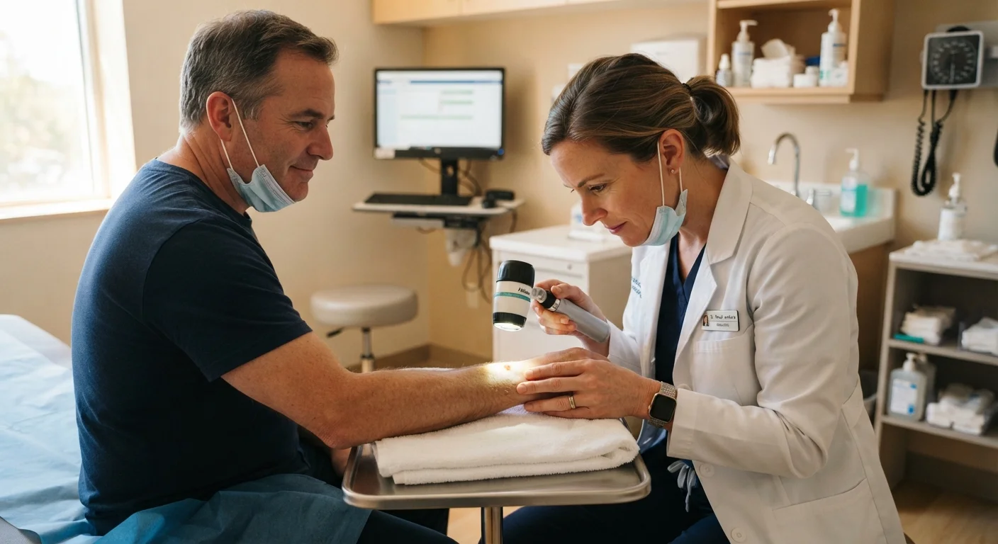
Any spot the clinician finds noteworthy is evaluated against diagnostic criteria and documented in your record. Many offices photograph suspicious lesions to enable comparison at future visits. The dermatologist may use a dermatoscope on specific areas for a closer, magnified view of pigmentation patterns and vascular structure.
Biopsy Decision (If Needed)
If a lesion raises enough concern, the dermatologist may recommend a biopsy during the same appointment or schedule one as a follow-up. A skin biopsy involves removing a small tissue sample under local anesthesia for laboratory analysis. This is the only definitive way to diagnose or rule out skin cancer. Most biopsies take only a few minutes and are minimally uncomfortable.
Post-Exam Counseling and Follow-Up Planning
At the close of the exam, the dermatologist will summarize findings, advise on any lesions being monitored, and recommend how frequently you should return based on your individual risk profile. They may also review sun-protective behaviors and monthly self-exam techniques so you can track changes between professional visits.
Understanding the Warning Signs Dermatologists Use
Dermatologists rely on systematic frameworks to evaluate spots and lesions consistently. The most widely used is the ABCDE criteria, applied primarily to moles and pigmented lesions:
- A — Asymmetry: One half does not mirror the other.
- B — Border: Edges are irregular, ragged, notched, or blurred.
- C — Color: Uneven distribution; multiple shades of brown, black, red, white, or blue.
- D — Diameter: Larger than roughly 6 mm (about the size of a pencil eraser), though smaller lesions can also be malignant.
- E — Evolution: Any change in size, shape, color, or a new symptom such as bleeding or itching.
For non-pigmented lesions — pink, pearly, or flesh-toned growths associated with basal cell and squamous cell carcinomas — dermatologists look for persistent sores that don't heal, raised or rolled edges, and unusual texture. A dermatoscope (a handheld magnifying device with polarized light) allows the clinician to examine subsurface structures not visible to the naked eye, improving diagnostic accuracy.
This targeted clinical approach is analogous to the systematic evaluation used in other preventive visits, such as those described in our guide on what actually happens during an annual physical exam.
This article is for general informational and educational purposes only and does not constitute medical advice. Always consult a qualified dermatologist or healthcare provider for personal guidance about skin health, risk assessment, or any concerning skin changes.