

Key Takeaways
- Mammograms use low-dose X-ray and are the primary screening tool recommended by major U.S. health organizations.
- Breast ultrasound uses sound waves, involves no radiation, and is typically used as a supplemental or diagnostic tool.
- Dense breast tissue can limit mammogram effectiveness; ultrasound may detect additional findings in these cases.
- Neither test replaces the other — they are often used together for a more complete picture.
- Always discuss your personal risk factors and screening schedule with a qualified healthcare provider.
Option A
Mammogram
The established, guideline-recommended standard for routine breast screening.
Best for: Women at average risk following standard screening schedules, typically starting at age 40 or 50 depending on clinical guidelines.
Option B
Breast Ultrasound
The flexible, radiation-free supplemental imaging tool.
Best for: Women with dense breast tissue, those who need follow-up after an abnormal mammogram, or individuals for whom radiation exposure is a concern.
If you are at average risk and following a routine screening schedule
Mammogram
Mammography is the only breast screening method with substantial evidence supporting reduced mortality from breast cancer in average-risk women and is endorsed by major U.S. guidelines.
If you have extremely dense breast tissue identified on a prior mammogram
Breast Ultrasound
Ultrasound can detect cancers in dense tissue that appear invisible on mammograms, and many U.S. states now require providers to notify patients of dense breast findings.
If you need to investigate a specific lump, cyst, or abnormal mammogram result
Breast Ultrasound
Ultrasound excels at characterizing whether a mass is solid or fluid-filled and is the preferred diagnostic follow-up for targeted evaluation.
If you are pregnant or cannot be exposed to ionizing radiation
Breast Ultrasound
Because ultrasound uses sound waves rather than X-rays, it carries no radiation risk and is generally considered safe for use during pregnancy.
If you are at elevated risk and want the most comprehensive screening
Mammogram
High-risk individuals are often screened with mammography plus MRI; ultrasound may be added when MRI is unavailable, but this decision should be guided by a physician.
How Each Imaging Method Works
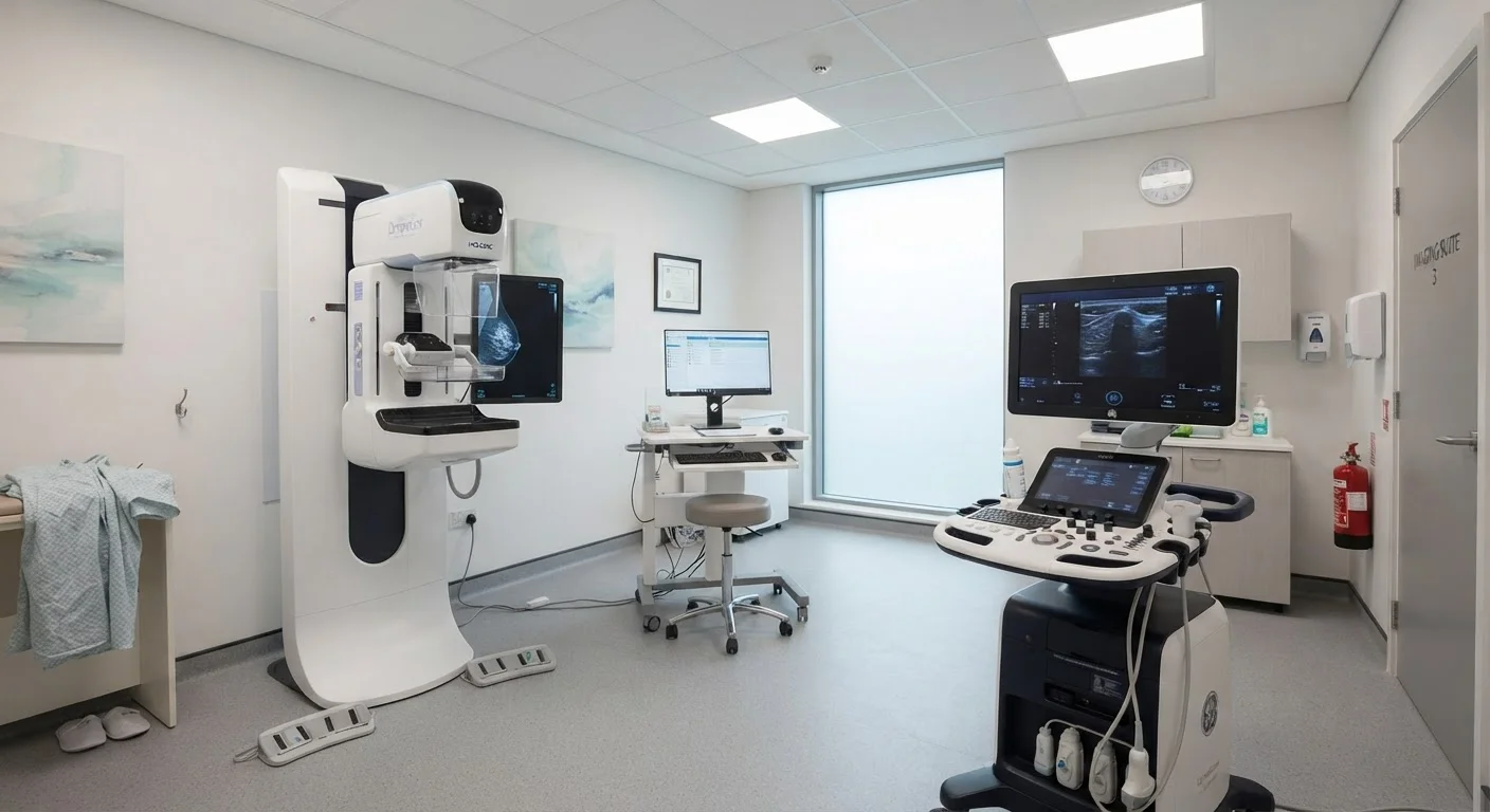
A mammogram is an X-ray of the breast. During the procedure, each breast is briefly compressed between two plates while a low-dose X-ray image is captured. The compression, though temporarily uncomfortable, improves image quality and reduces the radiation dose needed. Digital mammography — now standard in the U.S. — produces images that can be enhanced and analyzed on screen. 3D mammography (also called DBT) captures multiple thin-slice images to create a layered view, which can improve detection rates and reduce callbacks for additional imaging.
A breast ultrasound uses high-frequency sound waves emitted from a handheld device called a transducer. The transducer is moved across the skin's surface, and the reflected sound waves are translated into real-time images. No radiation is involved. Ultrasound is particularly sensitive at distinguishing between solid masses and fluid-filled cysts — a distinction mammograms cannot reliably make on their own.
| Criterion | Mammogram | Breast Ultrasound |
|---|---|---|
| Imaging technology | Low-dose X-ray | High-frequency sound waves |
| Radiation exposure | Minimal ionizing radiation | None |
| Primary use | Routine screening | Supplemental or diagnostic imaging |
| Dense breast tissue | Reduced sensitivity | Performs well |
| Detects microcalcifications | Yes | Rarely |
| Distinguishes cysts from solid masses | Limited | Highly effective |
| Guideline-recommended for routine screening | Yes (average-risk women) | No (supplemental only) |
| Safe during pregnancy | Generally avoided | Generally considered safe |
What Each Test Can — and Cannot — Detect
Mammograms are highly effective at identifying microcalcifications — tiny calcium deposits that can be early markers of certain breast cancers — as well as masses and architectural distortions within breast tissue. However, dense breast tissue (which appears white on a mammogram, just like tumors) can obscure findings. The American College of Radiology estimates that roughly 40% of women in the U.S. have dense breasts, which may reduce mammogram sensitivity.
Breast ultrasound does not reliably detect microcalcifications, which limits its use as a standalone screening tool. Where it excels is in dense tissue: studies have found that supplemental ultrasound screening in women with dense breasts can identify additional cancers not visible on mammography alone. Ultrasound is also the preferred modality for evaluating a palpable lump to determine whether it is a benign cyst or a solid mass requiring further workup.
Dense Breast Notification Laws
As of recent years, most U.S. states have enacted laws requiring mammography providers to inform patients when their mammogram reveals dense breast tissue. In 2023, the FDA updated national mammography quality standards to require that all patients receive this notification. If you have dense breasts, ask your provider whether supplemental screening may be appropriate for you.
Screening Guidelines and Who Should Get What
U.S. guidelines on mammography vary somewhat by organization, but most recommend that average-risk women begin routine mammograms between ages 40 and 50 and continue through at least age 74. The U.S. Preventive Services Task Force (USPSTF), the American Cancer Society, and the American College of Radiology each offer nuanced recommendations — speaking with your doctor helps determine the right schedule for you.
Breast ultrasound is not currently recommended as a standalone routine screening tool for average-risk women by major U.S. health organizations. Its role is supplemental: added to mammography for women with dense breasts, used diagnostically after an abnormal finding, or employed when mammography is contraindicated. Women at elevated risk — due to genetic mutations, strong family history, or prior chest radiation — may be advised to undergo additional imaging such as MRI, with ultrasound as a possible alternative when MRI is unavailable.
~40%
U.S. women with dense breast tissue
According to the American College of Radiology, approximately 40% of women who undergo mammography are found to have dense breasts, which can affect imaging sensitivity.
3–4 per 1,000
Additional cancers found with supplemental ultrasound
Research published in peer-reviewed radiology literature suggests supplemental ultrasound detects roughly 3–4 additional cancers per 1,000 women with dense breasts who had a negative mammogram.
This article is for general informational purposes only and does not constitute medical advice. Please consult a qualified healthcare provider to determine the screening approach appropriate for your individual health history and risk factors.